cECL Western Blot Kit
2023-10-22
RNase inhibitor, Murine
2023-10-22eECL Western Blot Kit
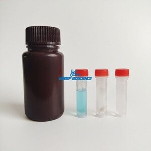
Product Number: WB049
Shipping and Storage
2-8 ℃, stored in dark
Components
| Component | WB049 50ml | WB049 250ml |
| eECL-A(Luminol Enhancer) | 25 ml | 125 ml |
| eECL-B(Peroxide) | 25 ml | 125 ml |
Description
The eECL Western Blot Kit is a highly sensitive and enhanced detection kit used in immunoblotting experiments in conjunction with horseradish peroxidase (HRP). This product is developed based on a new generation of enhanced chemiluminescent substrates, which undergo chemical reactions under the catalysis of HRP and emit light. It can be used to detect biomolecules such as proteins fixed on membranes. Its high sensitivity can detect PG level antigens, and the luminescent signal is strong and persistent. It can be detected using X-ray film exposure or chemiluminescence imaging equipment.
Note
- During contact with the membrane, please wear gloves and use clean equipment such as tweezers to avoid protein contamination and high background.
- Under dark conditions, the prepared chemiluminescence detection substrate working solution can be stably stored at room temperature for 8 hours. Sunshine or other strong light can affect the working fluid, so prolonged exposure to strong light should be avoided. Short term exposure to normal laboratory lighting does not affect the use of working fluids.
- Our company provides a variety of protein transfer membranes, blocking solutions, primary antibodies, enzyme-linked secondary antibodies, buffer solutions, etc. Please refer to our company website for details.
Protocol
- After the second antibody incubation is completed, wash the imprinting film thoroughly.
- According to the required amount, mix eECL-A and eECL-B in a 1:1 ratio and equal volume to prepare a chemiluminescence detection substrate working solution (approximately 1 ml of working solution is used for an 8 cm x 6 cm membrane).
- Discard the washing buffer and drop the luminescent substrate working solution onto the imprinting film, ensuring that the working solution covers the entire film. Incubate at room temperature for 3-5 minutes.
- Use a pipette to remove excess luminescent substrate working solution and place the imprint film between two clean plastic films. This process should be completed carefully to avoid the formation of bubbles between films.
- Expose X-ray film in a darkroom or place the film in a chemiluminescence imager and perform testing according to the instrument manual.
Schedule
| Problem | Reason | Resolution |
| Film inversion (White stripe, black background) | Excessive HRP in the system | Dilute HRP markers at least 10 times or more |
| Brown or yellow stripes appear on the membrane | ||
| Strong luminescence seen in the darkroom | ||
| The duration of the luminous signal is too short | ||
| Weak or no signal | Excessive HRP in the luminescent reaction system leads to rapid substrate consumption, resulting in rapid signal reduction | Dilute HRP markers at least 10 times |
| Insufficient antigen/antibody levels | Increase antigen/antibody usage | |
| Low protein transfer rate | Optimize transfer system | |
| High background | Excessive HRP in the system | Dilute HRP markers at least 10 times |
| Insufficient closure | Optimize closed programs | |
| Improper selection of sealing reagents | Choose another blocking reagent | |
| Insufficient flushing | Increase flushing time and frequency | |
| Overexposure | Reduce exposure time | |
| Antigen/antibody concentration too high | Reduce antigen/antibody usage concentration | |
| The protein bands are punctate | Protein transfer failure | Optimize the transfer process |
| Membrane imbalance | Handle the film according to the instructions | |
| There are bubbles between the film and film | Remove all bubbles before exposure | |
| Non specific bands appear (High background, short signal maintenance time) | There are too many HRPs in the system | Dilute HRP markers |
| Non specific bands appear (The background is clean and the signal maintenance time is normal) | Excessive dosage of primary antibody | Further dilution of primary antibody |
| SDS leads to non-specific binding | Avoid using SDS during the experiment |