1. How to determine the required nmol amount of primers for synthesis?
This depends on your experimental purpose. Taking a ~20-base primer as an example: for conventional PCR amplification, 5 nmol (approx. 1 OD) is recommended, which is sufficient for 200 standard 50 μl PCR reactions. For gene splicing or ligation after annealing, 5 nmol (approx. 1 OD) is also an appropriate choice.2. How to measure the OD value of primers?
The OD value of synthesized primers is measured using a UV spectrophotometer at a wavelength of 260 nm, with a quartz cuvette of 1 cm path length to determine the optical density of the solution. During measurement, the optical density should ideally be diluted to a range between 0.2 and 0.8. After the dry DNA powder is fully dissolved by vigorous shaking in a certain volume of water, take a portion and dilute it with 1 ml of water to measure the OD value. The OD value of the stock solution needs to be back-calculated based on the dilution factor. For instance, a simple way to verify if the amount of a 2 OD primer is accurate is as follows: add 1 ml of water, completely dissolve and mix thoroughly, take 100 μl, add 900 μl of water, and use a quartz cuvette with a 1 cm path length at a 260 nm wavelength; the absorbance reading should be 0.2.3. How to calculate primer concentration from the OD value?
Both GenScript's synthesis report and the primer tubes label indicate the OD value and molar amount. Primers are more stable when stored at high concentrations. Before dissolution, please verify whether the OD value on the synthesis report matches that on the primer label. If there is any discrepancy, please contact us immediately. We can track the actual yield through our production records.
According to international standards: 1 OD of dry primer powder is approximately 33 μg; the molar amount of the primer (μmol) = mass / molecular weight = (OD value × 33) / primer molecular weight.
For example, if you receive a tube of 2 OD primer with a molecular weight of 6565.3, the molar amount (μmol) = (2 × 33) / 6565.3 ≈ 0.010 μmol = 10 nmol.
If you need to reconstitute it into a 10 μM (= 10 pmol/μl) solution, simply add 1 ml of sterile ddH2O or 10 mM TE buffer (pH 7.5) and dissolve thoroughly.4. How is the molecular weight of primers (including modifications) determined?
The molecular weights of Oligos provided by GenScript are calculated using a precise algorithm.
Molecular weight calculation formula: MW = A × 313.21 + G × 329.21 + C × 289.18 + T × 304.2 + M × 301.2 + R × 321.21 + W × 308.71 + S × 309.2 + Y × 296.69 + K × 316.71 + V × 310.53 + H × 302.2 + D × 315.54 + B × 307.53 + N × 308.95 + 16 × Ns + molecular weight of modification groups - 61.96.
In the formula, Ns represents the number of phosphorothioate linkages, each adding 16 to the molecular weight; all other letters represent the count of the respective bases. For degenerate bases, the molecular weight is taken as the average of the corresponding bases, e.g., M = A/C = (313.21 + 289.18)/2. Common degenerate base codes: M=A/C, R=A/G, W=A/T, S=G/C, Y=C/T, K=G/T, V=A/G/C, H=A/C/T, D=A/G/T, B=G/C/T, N=A/G/C/T.
Table 3. Molecular Weights of Common Modification Groups
| Modification Group | Molecular Weight | Modification Group | Molecular Weight |
|---|---|---|---|
| 5'-Biotin | 447.11 | 3'-TAMARA | 623.60 |
| 5'-(6 FAM) | 537.46 | 3'-(6 FAM) | 569.46 |
| 5'-HEX | 744.13 | 3'-Amino Modifier C3 | 153.07 |
| 5'-TET | 675.24 | 3'-Amino Modifier C7 | 211.18 |
| 5'-Cy5 | 644.7 | 3'-Thiol Modifier C3 | 154.12 |
| 5'-Cy3 | 618.7 |
5. How should primers be stored?
Following synthesis, processing, and purification, primers are lyophilized into a flake-like substance. Lyophilized primers are highly stable and can be stored at -20°C for 2–3 years or even longer. Dissolved primers can be stored at -20°C for at least 6 months, though repeated freeze-thaw cycles must be avoided. If your experiments demand high reproducibility or the synthesized OD value is large, it is recommended to reconstitute the primers as a 100 μmol/L stock solution, aliquot them, and store them at -20°C. Before use, dilute the stock solution into a working solution (10 pmol/μl or 20 pmol/μl). Fluorescently modified primers must be protected from light.6. Will primers degrade if shipped at room temperature?
No, they will not degrade. Lyophilized primers remain stable at room temperature for at least two weeks. Since typical shipping times range from 1 to 3 days, the primers you receive will not experience degradation.7. How to dissolve primers?
Lyophilized primers have a highly porous and loose texture. Before opening the tube cap for dissolution, it is highly recommended to centrifuge at 3,000–4,000 rpm for 1 minute, or gently tap the tube vertically on the benchtop several times to collect the primer powder at the bottom, preventing powder loss upon opening. Add deionized sterile water or 10 mM Tris (pH 7.5) buffer based on the calculated volume, let it stand at room temperature for a few minutes, mix thoroughly by vortexing or pipetting, and centrifuge briefly to collect the solution at the bottom. Distilled water should generally be avoided for dissolving primers because some distilled water has a low pH (pH 4–5), under which conditions primers become unstable.
Our synthesis report specifies the required water volume to reconstitute each tube of primer to a concentration of 100 μmol/L (i.e., 100 pmol/μl). You may add an appropriate volume of nuclease-free double-distilled water (pH > 6.0) or TE buffer (pH 7.5–8.0) according to your experimental needs.8. Why do previously well-functioning reconstituted primers perform poorly after some time?
If the water used to dissolve the primers has a pH that is too low, or if it is contaminated with bacteria or nucleases, the primers will degrade. Failure to fully thaw and mix the solution before use can lead to non-uniform concentrations, resulting in inaccurate pipetting volumes. It is recommended to aliquot the primers, avoid repeated freeze-thaw cycles, and use 10 mM Tris (pH 7.5) buffer for dissolution. Another possibility is that the primers are fine, but the quality of the PCR materials—especially the template—is inconsistent with previous runs.9. How to test primer purity?
The most common laboratory method is PAGE (Polyacrylamide Gel Electrophoresis). This involves using a polyacrylamide gel supplemented with 7M urea. For primers with fewer than 12 bases, a 20% gel is used; for 12–60 bases, a 16% gel; and for more than 60 bases, a 12% gel. Take 0.2–0.5 OD of the primer, dissolve it in a saturated urea solution (or add urea powder directly to the primer solution until saturated), and heat-denature it before loading (95°C, 2 min). Urea serves both to denature the sample and to increase its density for easier loading. Electrophoresis is carried out at 600V for approximately 2–3 hours, after which the gel is removed and visualized on a fluorescent TLC plate under a UV lamp. The absence of minor bands beneath the main band indicates excellent purity. (Note: Incomplete denaturation can sometimes cause bands to appear above the main band, which are due to secondary structures of the primer.)
Additionally, analytical instrumentation can determine primer purity with greater accuracy:
- HPLC (High-Performance Liquid Chromatography): Separates components via gradient elution based on differences in adsorption strength. The mechanism relies on solute and solvent molecules competing for active sites on the adsorbent surface when the sample enters the column (reversed-phase columns are predominantly used today).
- CGE (Capillary Gel Electrophoresis): A liquid-phase separation technology driven by a high-voltage electric field that uses a capillary as the separation channel. It achieves separation based on differences in mobility and partitioning behavior among components, providing highly effective purity analysis. GenScript has adopted this as a major tool for primer quality control.
10. Is the primer purity acceptable if the OD260/OD280 ratio is less than 1.8?
The OD260/OD280 ratio cannot be used to evaluate primer purity. A low OD260/OD280 ratio is typically due to a high C/T content in the primer sequence. The table below lists the OD260/OD280 ratios of various 20-mer homopolymer primers, clearly demonstrating that the ratio is heavily dependent on base composition.
| Base Composition | OD260/OD280 |
|---|---|
| 5'-AAAAAAAAAAAAAAAAAAAA-3' | 2.50 |
| 5'-GGGGGGGGGGGGGGGGGGGG-3' | 1.85 |
| 5'-CCCCCCCCCCCCCCCCCCCC-3' | 1.15 |
| 5'-TTTTTTTTTTTTTTTTTTTT-3' | 1.14 |
| 5'-AAAAAGGGGGTTTTTCCCCC-3' | 1.66 |
11. Why does EB staining intensity vary for the same OD amount during PAGE analysis?
EB (Ethidium Bromide) staining is typically used to quantify double-stranded DNA (such as plasmid DNA) because EB fluoresces by intercalating between the bases of a double helix. Synthesized single-stranded DNA oligos, however, can only be stained by EB if they fold back on themselves to form local hairpin loops or inter-chain partial double helices. Because of variations in sequence composition, different primers have different propensities to form secondary structures, leading to variations in EB staining intensity. For example, Oligo(dT) does not form secondary structures, resulting in very poor EB staining. Therefore, EB staining should not be used for single-stranded DNA quantification; instead, measurements should be performed using a UV spectrophotometer.12. During PAGE, why do Oligo DNAs of the exact same length migrate to different positions?
This phenomenon is more pronounced in shorter Oligo DNAs; longer oligos show less variation. There are two main reasons for this:
- Differences in A, G, C, and T composition alter the migration rate.
- Differences in the three-dimensional conformation of the DNA alter the migration rate.
13. Can agarose gel electrophoresis be used to analyze synthesized primers?
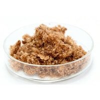
Denaturing PAGE must be used for primer electrophoresis. Because primers are single-stranded DNA, they easily form complex three-dimensional structures. Consequently, agarose gel electrophoresis frequently yields multiple bands or no bands at all, making it entirely unsuitable for quantification.14. Why do lyophilized primers sometimes appear yellowish-brown? Is this the natural color of DNA?
Synthesized primers can appear yellowish-brown, white, or transparent. This variation depends on the base composition and the synthesis manufacturing process. Sequences rich in A and G bases, as well as primers with high OD values, often exhibit a yellowish-brown tint. This coloration will not have any impact on your experimental results.15. If a PCR amplification fails, is it related to the primers?
In most cases, no. The continuous development of diverse PCR technologies and various thermostable polymerases is specifically intended to overcome challenges like non-amplification or low amplification efficiency. For example, nested PCR is designed to amplify gene fragments with very low copy numbers. Amplifying repetitive sequences or fragments with high GC content typically requires specialized amplification strategies.
When amplification fails, the following two scenarios are most common:
- RT-PCR: Many genes are inherently difficult to amplify via conventional RT-PCR. The success of RT-PCR relies heavily on the quality of the RNA used in the RT reaction and the abundance of the target gene in the specific tissue or cell type.
- Amplification from Genomic DNA: Generally, genes exist as single copies within the genome, meaning genomic DNA template amounts must be strictly controlled. Excessively high amounts of genomic DNA can disrupt the Mg2+ concentration and pH of the reaction system.
If you have ruled out other factors and suspect an issue with the primer, please provide us with the specific details. We will evaluate the issue and offer a suitable solution.16. What should I do if there is no target band when using synthesized primers in a PCR?
PCR failure can stem from numerous factors. Consider troubleshooting the following aspects:
- Are the primers and template properly paired, and what is their degree of homology?
- Do the primers themselves form stable secondary structures, or do primer-dimers/higher-order structures form between the two primers?
- Are the PCR reagents functioning properly?
- Is the thermal cycler operating correctly?
- Are the PCR cycling conditions optimized?
If all parameters check out and the problem persists, we will resynthesize the primers once free of charge.17. Does strong non-specific amplification indicate primer contamination?
No. Sequencing analysis of non-specific bands typically reveals at least one of the primer sequences at both ends of these fragments. Therefore, non-specific amplification is generally caused by template contamination (such as genomic DNA contamination in RNA samples) or sub-optimal PCR conditions.18. How to anneal two complementary single strands into double-stranded DNA?
Dissolve the primers in annealing buffer (10 mM Tris, pH 7.5–8.0, 50 mM NaCl, 1 mM EDTA). Mix equimolar amounts of the complementary primers, keeping the total volume below 500 μl. Heat the mixture to 95°C for 2 minutes, then allow it to cool slowly to room temperature (below 30°C). The annealed product can be stored at 4°C for subsequent use.19. Why do annealed primer fragments fail to ligate into the vector?
If the primers used in the ligation reaction lack a 5' phosphate group, ligation efficiency will be significantly lower, though not entirely impossible. If phosphorylated products still fail to ligate, verify the digestion efficiency of your vector and double-check your primer design. If both are correct, consider optimizing the primer annealing conditions and the ligation reaction system.
Primer Synthesis and Purification
Primer Synthesis, Storage, and Usage
Primer Design and Modification